- Doctors recommend bitewing X-rays once a year to detect cavities and decay between the teeth. You may need a periapical radiograph to diagnose fractures, cracks, and abscesses.
- A Full-Mouth-X-ray (FMX) includes 16 to 18 images to view the entire dentition and incoming teeth. Occlusal X-rays help identify the need for caries treatment.
- Most dental insurance plans cover the cost of X-rays, and prices typically range from $25 to $750 depending on the type and number of images needed.
Need a dental x-ray this week? Use Authority Dental to find emergency dental clinic near you.
Dental X-rays typically cost $25 to $750 depending on the type. Learn the cost of bitewing, panoramic, FMX, and CBCT X-rays, how often you need them, and what insurance usually covers.
Intraoral X-rays
Intraoral means inside the mouth. That is precisely where the film goes when these X-rays are taken. Intraoral radiographs are the most common dental exam and provide the most detail.
Bitewing X-ray
Picture by Authority Dental under CC 2.0 license
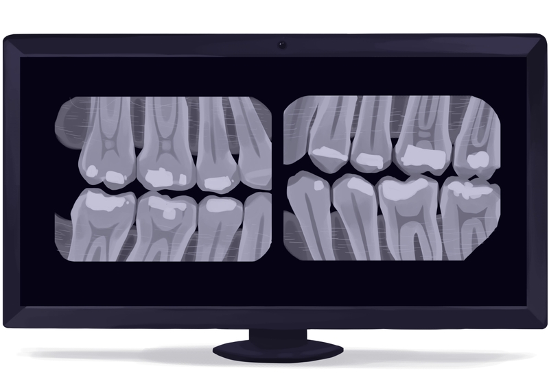
A bitewing is performed once a year, unless necessary, before a particular treatment. It is sometimes referred to as a "check-up X-ray. That is because it is swift and easy, and because it provides the dental professional with a great deal of information, especially about tooth decay and between-teeth decay.
Harry Lee, DMD, explains: "The Bitewing is what we take routinely, typically once a year, and it is invaluable for spotting minor issues between the teeth. From conversations with colleagues, we all agree that if we skip this check-up X-ray, we inevitably miss small interproximal cavities that become significant, painful problems six months later."
"It allows us to catch decay when it is still small and treatable with a simple filling, saving the patient from a future root canal," he emphasizes.
Tooth decay occurring on the inside of your tooth or between your teeth can be easily diagnosed. Changes in bone thickness, signs of gum disease, and tooth decay will also appear on the image.
Bone loss can be caused by tooth loss or tooth decay, as well as by chronic periodontitis, a preventable issue resulting from poor oral hygiene. Bone loss can affect anyone, regardless of age. However, if you want healthy teeth, you can prevent them not only with good oral hygiene but also with regular dental check-ups, which most dental insurance plans cover.
With specific dental imaging technology, dentists can spot bone loss in its early stages. Moreover, while insurance coverage of such exams may vary from one company to another, it is often convenient to have your teeth and mouth conditions examined by a professional dentist.
A bitewing captures a small section of the lower and upper teeth. If you are not focusing on a problem area, multiple can be taken for a general overview. Additional radiographs are usually less expensive than the first one. In some cases, they may be taken at no additional charge.
The name stems from the shape of the device you put into your mouth. You bite down on a wing-shaped piece of film. The dentist or hygienist leaves the room for each snapshot, and you might wear a protective bib.
Related procedures include dental exams, professional teeth cleaning, cavity filling, and cosmetic treatments.
Periapical X-ray
Picture by Authority Dental under CC 2.0 license
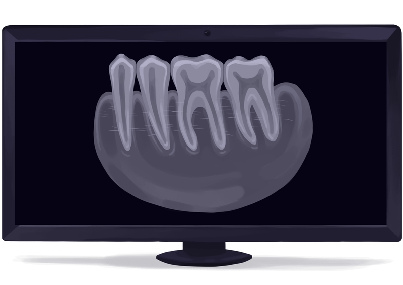
A periapical X-ray shows a small section of the mouth, concentrating on one or two teeth. The entire tooth from the crown to the tip of the root (below the gums) will be visible. It is usually performed 1-2 times a year.
It may help diagnose issues you did not know you had, such as impacted teeth, fractures, cracks, and abscesses. It is most often taken when the patient experiences acute pain. This could be undiagnosed pain or pain that is suspected to be caused by failure of prior treatment.
The dental professional will leave the room. You should wear a protective bib, one covering your thyroid gland at least. The film will usually be mounted on a metal rod with a ring. You will have to bite down on the device. This will help it stay still.
You will need more than one picture. Additional radiographs are usually less expensive than the first.
Periapical X-rays are taken when you come in for teeth whitening, SRP, RCT, veneers, crown placement, and tooth extraction.

FMX
Picture by Authority Dental under CC 2.0 license
Sometimes the dental hygienist or dentist decides that, rather than taking individual periapical or bitewing X-rays, an FMX is needed.
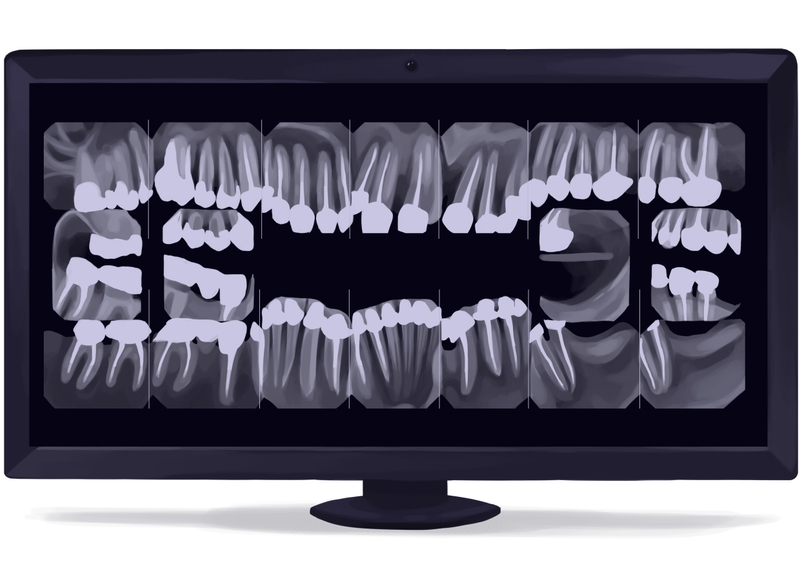
FMX means a full-mouth series of X-rays. It includes a set of 16-18 images to observe the complete set of teeth, incoming teeth, and the surrounding tissue. These usually include four bitewings and 12-14 periapicals.

The radiographs will show the structure of your entire mouth, including details of individual teeth. The jaw will be presented from both sides. This will give the dental professional a good idea of the general structure of your mouth.
They can detect abnormalities in the mouth's structure, including decay, gum disease, and abscesses.
The procedures will not differ from those described above. Some offices require you to have an FMX if you are a new patient. For established patients, four bitewings and two periapicals (of the front teeth) are usually taken every 6-12 months.
Occlusal X-ray
Picture by Authority Dental under CC 2.0 license
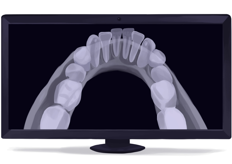
This type of X-ray concentrates on tooth development. Each radiograph shows an entire arch, upper or lower. An occlusal X-ray is most commonly used to detect the need for a cavity filling or to assess orthodontic work.
It is mainly performed in children experiencing problems with unusual tooth alignment. It also captures unerupted teeth, or teeth that are erupting in unexpected places. The image shows the roof or floor of the mouth.
Caries below the gumline, fractures in the jawbone, abscesses, and lodged foreign objects can be easily spotted.
Extraoral X-rays
For the following three types of X-rays, the film remains outside the mouth. These show the teeth and the surrounding structures, such as the jaw and skull.
Panoramic X-ray
Picture by Authority Dental under CC 2.0 license
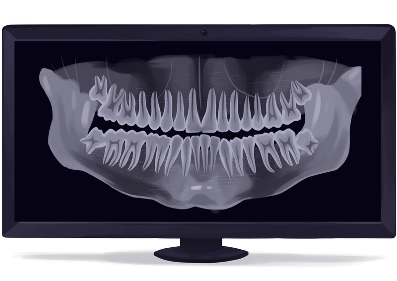
A panoramic X-ray is a common procedure at the dental office. It provides a big-picture view of the relationship between the jaws and teeth, as well as the airways, such as the nasal cavities and sinuses. This is a non-invasive test that is the way to go if you are unsure which tooth is hurting.
It requires a little preparation. You will have to remove your jewelry, glasses, and other metal objects. You will be given a lead apron to protect the rest of your body from radiation exposure.
You might have to stay standing for this X-ray. Your head and chin will be placed in a device that will help you remain motionless. This is vital, as any movement could distort the radiograph.
Even though the device will rotate around you, all teeth will be shown on a flat image. Both arches, along with surrounding structures and tissues, will be visible.

You may have it if you choose a procedure that requires a comprehensive dental evaluation, such as implant placement. Many orthodontists also like to take a panoramic X-ray of the mouth before performing any orthodontic work.
Other than before major procedures, it can be done every 3-5 years.
Cephalometric projections
Picture by Authority Dental under CC 2.0 license
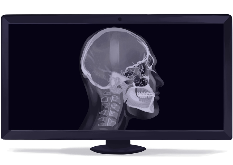
A “ceph,” as it is sometimes referred to, is usually taken before and after orthodontic treatment. This includes not only traditional braces but also clear aligners such as Invisalign.
This X-ray concentrates on the patient’s profile and helps predict the outcome of tooth movement. Exposure takes approximately ten seconds, and the image is developed in about five to six minutes. It will be two-dimensional.
The dental professional will use tracing paper to “trace the ceph. The movement of the dentition and its growth patterns can be calculated.
Cone-beam computed tomography (CBCT)
Picture by Authority Dental under CC 2.0 license
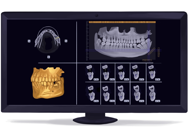
A cone-beam CT is often confused with a medical CT scan, though it delivers much less radiation. It provides considerably more details than other types of dental X-ray, as the image produced is 3D.
It is used when other types prove insufficient, before oral surgery, or in invasive RCTs. During the procedure, a cone-shaped beam will move slowly around your head. You will have to remain motionless for the shot to be precise.
Three radiographs are captured in a single scan. Those pictures, or “views, are then combined to produce a three-dimensional image. The dentist will be able to assess space and internal structures in the mouth. The teeth, soft tissues, nerve pathways, and bone structure will all be visible.
The dentist will also be able to see which direction the roots grow in. This makes oral surgery less invasive; it is easy to see where the incisions have to be made.
Cone beam X-rays are the most detailed, accurate, and diagnostic form of radiograph. This type is invaluable for implant placement, wisdom tooth extraction, and root canal treatment. Some complications, such as permanent nerve damage, can be more easily avoided if this X-ray is done.
Digital dental radiographs
Advances in dental technology and medical science have significantly impacted routine dental x-rays, dental procedures, and overall oral health.
Digital X-rays expose the patient to significantly less radiation than traditional X-ray film radiographs. Safety is the most important reason for using this type of dental imaging. Since the radiation level is lower, this allows for more frequent routine dental X-rays to help prevent oral disease and promote overall dental health.
Digital radiography is a new technology that might one day replace traditional X-ray film. It is not a type of radiograph but a different means of taking one. You can, for example, take a bitewing this way.
The advantage is that images are processed immediately so that they can be viewed straight away. These images are editable; the technician can adjust contrast and deduce more information from the radiograph. There is no real difference in terms of price for the patient.
One of the most significant disadvantages is that some sensors are larger and bulkier than traditional ones. This means it is less comfortable for the patient and cannot be easily disinfected. The device is covered in disposable plastic for infection control.
Digital X-rays expose the patient to significantly less radiation than traditional X-ray film radiographs. Besides, this type of radiography is commonly used to detect tumors. There are many types of tumors, including dental tumors, which often develop under the soft tissue of the jaw. These tumors can be benign or malignant and can be detected on these radiographs.
Cost of dental X-rays explained
Dental X-rays near you vary from $25 to $750. The type of dental X-ray you need, and, of course, how many, are what impact the price most. A dental professional is the one who can accurately diagnose what kind of treatment is appropriate in your case.
The cost of intraoral and extraoral X-rays can be very different.
| INTRAORAL X-RAYS TYPE | AVERAGE COST | COST RANCE |
|---|---|---|
| Bitewing | $35 | $25-$50 |
| Periapical | $35 | $25-$50 |
| FMX | $150 | $100-$300 |
| Occlusal | $50 | $25-$100 |
And prices for extraoral X-rays:
| EXTRAORAL X-RAYS TYPE | AVERAGE COST | COST RANCE |
|---|---|---|
| Panoramic | $130 | $100-$250 |
| Ceph | $150 | $70-$300 |
| Cone-beam CT | $350 | $150-$750 |
In general, extraoral X-rays are usually more expensive. This is because they require equipment that costs a lot more. Moreover, small offices may not have the necessary tools, and you may be sent elsewhere.
If that is the case, you should be prepared for a higher fee. Dental offices do this to avoid an influx of patients who are just going to do minor procedures instead of complete treatment.
Are dental X-rays safe?
Generally, there are many misconceptions about radiography. The radiation produced during an X-ray is very small, comparable to the amount you would experience during a sunny day. The exposure from four bitewings is comparable to a two-hour airplane ride.
What is more, the benefits of early detection of any issues outweigh the risk of a minimal amount of radiation. There is also no reason to worry about children receiving X-rays. Radiography can be safely used in pediatric care.
One of the most significant contraindications to X-rays is pregnancy. Still, accommodation can be arranged if necessary. For example, if a woman becomes pregnant during long-term treatment, she and the baby will be protected by a heavy-duty leaded apron and a thyroid collar.

FAQ
How often do you need dental X-rays?
You should get a bitewing twice a year. It is convenient to do this during your bi-annual check-ups. An intraoral periapical should be done when you are a new patient to an office.
Other than that, X-rays are not routine procedures. They are done instead when something unexpected happens or when you are considering an invasive treatment. Radiographs can also be very useful for diagnosing dental problems.
How much radiation is there in dental X-rays?
Trace amounts of radiation are emitted by practically everything. Yes, even in everyday objects like bananas or cosmetic products. Dental X-rays produce more, but still less, than, let us say, a flight from New York to Los Angeles. Procedures like mammograms and abdominal CT scans are a lot more radioactive.
Can you get a dental X-ray during pregnancy?
Dental X-rays while pregnant are not prohibited. Taking these images is not recommended unless necessary, however. In such cases, protection in the form of a lead apron can be provided.
Are dental X-rays painful?
No. X-rays shine light onto parts of your body to produce an image; they do not touch you at all. You will not feel any pain or discomfort while one is being taken.
How to read a dental X-ray?
Unfortunately, reading X-rays takes years to master. You should rely on your dentist to decipher it. To make things simple, the white areas are your hard tissues (teeth, jaws, and ligaments), and the dark areas are the soft parts of your mouth, such as your gums and cheeks. Special equipment also allows measuring the distance between specific points in the image.
Does insurance cover dental X-rays?
Routine bitewings or even FMX will likely be covered, as they are considered preventive care. There is a chance that you will be reimbursed with no deductible. Thanks to their diagnostic nature, X-rays can also be paid for by insurance when taken to aid therapeutic or cosmetic treatment.
Additionally, you can consider signing up for a dental plan. They have no yearly maximums, waiting periods, or paperwork. In fact, they will not need an X-ray to give you a discount on any procedure. Reductions reach 60%.
Jason Chong, DDS
Bitewings are recommended every other cleaning visit, or every twelve months to evaluate for caries and to identify subgingival calculus.